Macular degeneration examination through fundoscopy: Insights on findings and patient expectations
Fundoscopy, also known as ophthalmoscopy, is a vital diagnostic method used by eye doctors to check for signs of Age-Related Macular Degeneration (AMD). This common eye condition primarily affects older adults and can lead to significant vision loss.
During a fundoscopy, the eye doctor examines each eye separately. The person will sit in a chair, looking straight ahead, and remove their glasses but not their contact lenses. The doctor will dim the lights in the room for the procedure and may use eye drops to dilate the person's pupils, which may take 15 to 20 minutes to dilate fully.
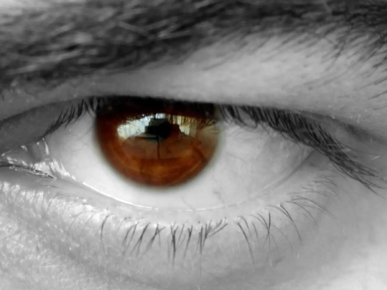
The dilated pupils allow the doctor to see the fundus, the inside back surface of the eye that includes the retina, macula, optic disc, fovea, and blood vessels. This comprehensive view is crucial in detecting early signs of AMD.
In AMD, a person may experience pigment changes and whitish fatty deposits under the retina called drusen. In its more severe form, wet AMD, abnormal blood vessels grow under the retina, which can lead to a variety of complications such as a gray-green membrane or thickening under the retina, blood, fat, or fluid under the retina, raised areas of the retina, bleeding, tears or rips, and scarring.
It's important to note that people with risk factors for AMD, such as smoking, a family history of the disease, and being over 50 years old, will likely need more regular check-ups. The American Academy of Ophthalmology recommends people age 40 and older have a baseline eye exam and then follow the recommended schedule for regular comprehensive, dilated eye exams.
The examination itself can be either indirect or direct. In indirect fundoscopy, the doctor uses a lens and light source to view the eye from a distance. Direct fundoscopy involves the use of a handheld ophthalmoscope to directly view the eye.
Following the examination, the person may experience blurry vision and sensitivity to light, so they should not drive for a few hours afterward. Additionally, the person may find it difficult to focus their eyes for up to 12 hours after fundoscopy.
Fundoscopy is just one of several tests doctors use to check for signs of AMD. If AMD is detected, early treatment can significantly slow its progression and help preserve vision. The American Academy of Ophthalmology provides resources to find an ophthalmologist in your area.
Regular comprehensive, dilated eye exams are essential for older adults, as they help in the early detection and treatment of AMD and other eye conditions. By staying vigilant and following the recommended exam schedule, individuals can help maintain their vision health and quality of life.
Read also:
- Nightly sweat episodes linked to GERD: Crucial insights explained
- Antitussives: List of Examples, Functions, Adverse Reactions, and Additional Details
- Asthma Diagnosis: Exploring FeNO Tests and Related Treatments
- Unfortunate Financial Disarray for a Family from California After an Expensive Emergency Room Visit with Their Burned Infant


